About Atrix
Lorem ipsum dolor amet consectetur adipisicing sed do eiusmod tempor incididunt labore magna aliqua enim minim veniam nostrud exercitation aboris nis aliquip exeo.
Find Us Our Location
- 629 12th St, Modesto, CA 95354 United States
- atrixmain@gmail.com
- +1 (230)-456-155-23
Blog Standard
The Role of Phosphorylation in Post-Translational Modification: Activating Akt/PKB
Clear cell renal cell carcinoma (ccRCC) is the most prevalent and aggressive subtype of cell renal cell carcinoma, presenting significant therapeutic challenges due to its radioresistance, chemoresistance, and genetic variability. With a five-year mortality rate of 40%, there is an urgent need for reliable prognostic biomarkers to facilitate personalized treatment approaches. Recent research suggests that monitoring oncoprotein activation states, specifically Akt/PKB (Protein Kinase B) and STAT3 activation, using QF-Pro® technology may offer a promising diagnostic tool with prognostic significance.
Current Limitations in ccRCC Biomarker Assessment
Traditional diagnostic techniques primarily rely on immunohistochemistry (IHC) analysis to evaluate protein expression levels. While effective, IHC analysis fails to provide insights into protein activation states and their functional relevance in disease progression. QF-Pro® technology overcomes these limitations by enabling biomarker analysis of oncoprotein activation through fluorophore-labelled antibodies, providing a more detailed and reliable analysis of biomarker functionality in cell renal cell carcinoma.
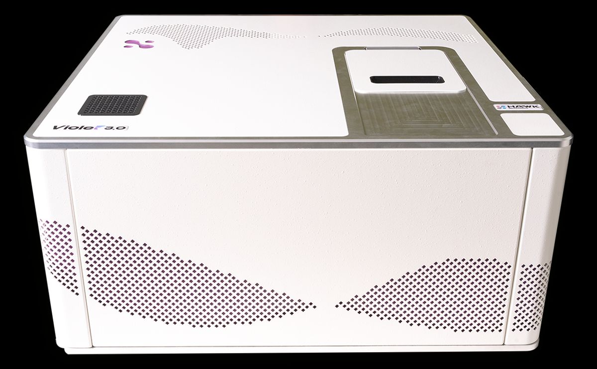
Hawk Biosystems Violet 3.0, integrated with QF-Pro® Technology
QF-Pro® Technology and Methodology
Principles of QF-Pro®
QF-Pro® analysis is based on Förster Resonance Energy Transfer (FRET)-Fluorescence Lifetime Imaging Microscopy (FRET-FLIM), a non-radiative energy transfer technique occurring between fluorophores when within a 1-10 nanometer proximity. This two-site amplified FRET-FLIM approach enhances specificity by reducing false positives and allowing for precise quantitative measurements of protein kinase B (Akt/PKB activation).
In this study, QF-Pro® technology was used to assess Akt/PKB activation and STAT3 activation in ccRCC samples by targeting phosphorylation-specific antibodies at key activation sites. This technique enables real-time visualization of oncoprotein activation, offering a critical advantage over traditional methodologies.
Sample Preparation and Analysis
Formalin-fixed, paraffin-embedded (FFPE) tissue microarrays (TMAs) from primary and metastatic ccRCC samples were analyzed. Secondary QF-Pro® probes tagged with ATTO 488 (donor) and Alexa594 (acceptor) fluorophores were used to detect activation state scores for each patient sample.
Results
Akt/PKB Activation Upon EGF Stimulation
Akt/PKB activation dynamics were assessed using growth factor (EGF) stimulation. QF-Pro® activation maps revealed marked differences between basal and stimulated conditions.
- In basal conditions, cells exhibited predominantly blue signals, indicating minimal Akt phosphorylation.
- After 10 minutes of EGF stimulation, cells transitioned to green, yellow, and orange, reflecting a progressive increase in Akt phosphorylation at the plasma membrane.
- Quantitative measurements confirmed a significant increase in Akt phosphorylation over time, with peak activation observed at 10 minutes post-stimulation.

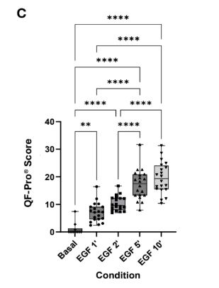
Figure 1. A) Illustration of QF-Pro® assay for detecting Akt/PKB activation. B) Representative QF-Pro® maps show minimal Akt/PKB activation under basal conditions and increased phosphorylation upon EGF stimulation for 10 minutes. C) Quantitative QF-Pro® scores across different EGF treatment time points demonstrate a significant increase in activation state with longer stimulation times (p < 0.001).
Akt/PKB Activation in ccRCC Progression
QF-Pro® analysis of renal control tissues, primary tumors, and metastatic ccRCC samples demonstrated clear differences in Akt/PKB activation states:
- Renal control tissues showed minimal activation (predominantly green QF-Pro® maps).
- Primary tumor samples exhibited a mild increase in activation.
- Metastatic samples displayed high phosphorylation levels (yellow and red QF-Pro® maps), indicating enhanced Akt/PKB activation during ccRCC progression.
These results highlight Akt/PKB signaling pathway as a potential biomarker for assessing tumor progression and aggressiveness in cell renal cell carcinoma.
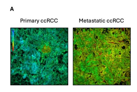
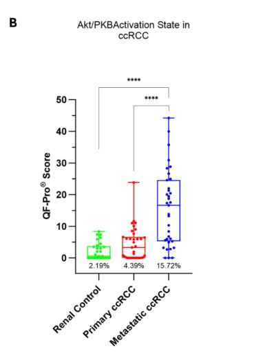
Figure 2. A) Representative fluorescence lifetime images, primary ccRCC tissue shows moderate Akt/PKB activation (blue/green) and metastatic ccRCC tissue exhibits increased activation (yellow/red). B) QF-Pro® Score demonstrated a significant difference between non-cancerous, primary, and metastatic samples (p < 0.001), confirming that Akt activation state, rather than mere expression levels, correlates with clinical prognosis in ccRCC patients.
Prognostic Precision of QF-Pro® vs. IHC
QF-Pro® technology demonstrated superior prognostic accuracy compared to IHC analysis, particularly in assessing Akt/PKB activation states:
- Kaplan-Meier survival analysis showed that patients with higher Akt activation (upper quartile) exhibited poorer survival outcomes.
- Conversely, IHC-based phosphorylation (Threonine 308) Akt expression failed to correlate with survival, highlighting its lower prognostic reliability.
- The limitations of one-site IHC assays in distinguishing activation states emphasize the need for functional biomarker analysis, which QF-Pro® technology uniquely provides.
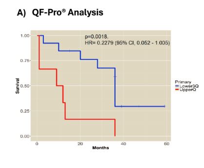

Figure 3. Akt/PKB activation state correlates with poor overall survival in ccRCC. Kaplan-Meier survival outcomes related to PKB/Akt activation as determined by QF-Pro® (A) or by conventional IHC (B).
A) QF-Pro® analysis revealed significantly poorer survival for patients in the upper quartile compared to the lower three quartiles.
B) No significant survival difference was observed using conventional IHC.
STAT3 Activation Dynamics
STAT3 activation (Tyr705 phosphorylation) was assessed across renal control tissues, primary ccRCC tumors, and metastatic samples:
- Renal control tissues exhibited low STAT3 activation.
- Primary ccRCC samples displayed a modest but statistically significant increase.
- Metastatic tumors showed a marked elevation in STAT3 activation, emphasizing its role in tumor progression and aggressiveness.
These findings suggest STAT3 activation as a potential biomarker for ccRCC prognosis, correlating increased activation with advanced disease stages and poor patient outcomes.
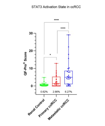
Figure 4. Tyr705 activation is higher in metastatic ccRCC tumors in FFPE TMAs. Box and Whisker plots show the QF-Pro® scores of the three groups. Activation of Tyr705 is higher in metastatic cores than in primary cores. Both groups are significantly higher than the non-cancerous renal control tissue (p <0.001).
Conclusion
QF-Pro® technology represents a significant advancement in biomarker analysis by measuring protein activation states, rather than just expression levels. This marks a paradigm shift from traditional IHC analysis, which is limited in detecting post-translational modifications (PTM).
- By focusing on phosphorylation (Threonine 308), QF-Pro® enhances specificity and sensitivity, offering critical insights into tumor progression.
- In cell renal cell carcinoma, QF-Pro® technology provides clinically relevant prognostic information, linking biomarker activation to tumor evolution and patient survival.
- By integrating multiple biomarkers, QF-Pro® strengthens risk stratification and supports personalized treatment approaches.
With its ability to measure functional protein states and monitor therapeutic responses, QF-Pro® analysis overcomes the limitations of conventional diagnostic methods, paving the way for more precise and individualized cancer care in ccRCC.
Inkarp Instruments is a trusted distributor and service provider of Hawk Biosystems products in India, offering innovative scientific solutions. With a focus on quality and reliability, we equip researchers across the country with advanced products and dedicated support.
References
1. Miles J, Applebee CJ, Leboucher P, et al. Time resolved amplified FRET identifies protein kinase B activation state as a marker for poor prognosis in clear cell renal cell carcinoma. BBA Clin. 2017;8:97-102. doi:10.1016/j.bbacli.2017.10.002
2. Veeriah S, Leboucher P, de Naurois J, et al. High-throughput time-resolved FRET reveals Akt/PKB activation as a poor prognostic marker in breast cancer. Cancer Res. 2014;74(18):4983-4995. doi:10.1158/0008-5472.CAN-13-3382